|
Feature and Characteristics of
the
MDI
detector
The MDI detector is a
kind of microanalysis apparatus
made in U.S.A. The MDI detector
uses color camera shots to collect
pictures whose resolution reaches
800×600 by means of the
systematic biological microscope
with high magnification times. The
detector can synchronously
transmit the linearly zoomed
medical images and magnifies the
object 10000
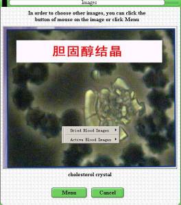
times. In the clear-cut ultramicro
camera field, the observer can,
through the monitor, observe
various blood configurations
including the roving glycerol, the
blue shiny cholesterin, the brown
uric acid crystals,
atherosclerosis clots and lipid
clots in the fresh blood sample.
Through observing the dry blood
sample, the MDI detector can
directly reflect the patients'
organ damages, degradation of the
arthrosis, blood oxidation (damage
of the freeradicle), cerebral
ischemia and tumors etc. It can
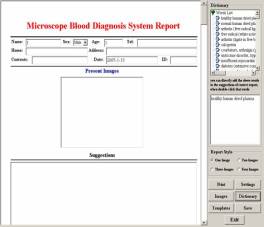
also connect with the computer
through LG special medical image
acquisition to realize real time
acquisition,labeling,
storage and archiving.
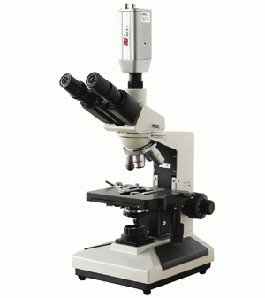
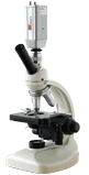
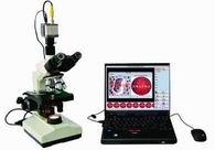
Top-grade
Convenient Type
Economic
Type
(Three
oculars)
(Double
oculars)
Applicability
of the MDI detector:
The
MDI detector can help detect
different types of sub-healthy
symptoms and assist analysis and
diagnosis of them ,such as:the
erythrocyte appearing to be
lemon-shape, money-cluster shape
or in bulk, gigantocyte(or
microcyte), target-shape,
bottle-top C type or Y shape,
echinosis, brittleness of the
erythrocyte, aggregation of the
thrombocyte, aleukocytosis
(<3.5×10/L) and leukocytosis
(>10×10/L) etc. There are many
causes that lead to those
symptoms, such as : weak
immunologic function, mycotic
infection, dyspepsia or
malabsorption, malnutrition,
vitamin deficiency, weak born
marrow function and allergic
reaction etc. Some might be caused
by circulation of toxic substances
(aggregation of thrombocyte,
needles, chyle grain, bioblast and
atheoscleorsis)
Parts
of the MDI detector apparatus:
The
biological microscope is made up
of: single-ocular/ binocular tube
lens cone, ocular , object lens
and converter, focus
adjustment mechanism,moving
objective table , base, lighting
system, 14″color monitor which
has more than 600 lines
distinguishability in the center
and can display high-quality image
on the 14″ screen. The color
monitor can accept the standard
NTSC/PAL color input signal.
Notes
of the MDI detector:
The
monitor can be replaced by TV or
computer screen.
Note for adjusting the TV set:
Adjust the abstract to the maximum
and the brightness to more than 10
and the chroma as high as
possible.
Note for using the computer:
Setting of the collection card:
video standards: PAL-D,
brightness:5000,
contrast:5000,chroma:5000,saturation:10000,frame
frequency:
29.970,color
space/compression:RGB24,set
the output power according to
need. The computer should be
equipped with high-quality show
card and high-contrast display to
have better display performance.
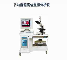
MDI connected with desk computer
MDI
connected with laptop

MDI
connected with TV set
|